The core of my service focuses on utilizing medical image processing algorithms, some of which are based on artificial intelligence methods. Thus, the outcome of my work is the generation of the following images:
Gastroenterology
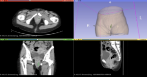
- Perianal Fistula
Perianal fistula is a research area that receives very little attention, even though thousands of patients are affected by it.
- Anal Abscess
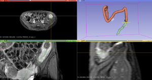
- Bowel Wall Thickening in MRI
Abnormal increase in the thickness of the gastrointestinal tract’s walls.
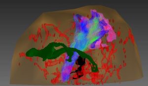
- Expanded Anatomy of Female Pelvis: Organ Prolapse, Perianal Fistulas, Muscles, Rectum and Urinary Bladder
Internal obturator muscle (purple), levator ani muscle (green), rectum (blue), perianal fistulas (yellow), gluteus maximus (orange), and urinary bladder (light yellow).
- Virtual Colonoscopy: Diverticulosis and Thickening
Neurosurgery and Orthopedics
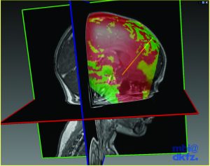
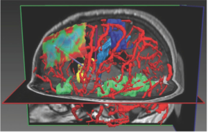
- Color-Coded Neuro Surgical Planning
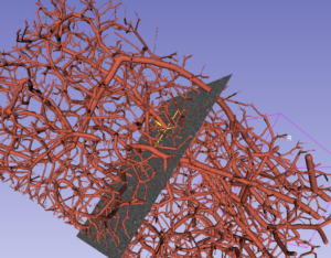
Here are the images I created during my thesis research. The first set showcases segmented structures, with colors representing distinct elements such as blood vessels in red, motor fMRI in dark purple, sensory fMRI in blue, vision fMRI in brown, vision fibers in yellow, and motor fibers in light blue. The second set displays a color-coded head surface, where green indicates low risk, yellow denotes medium risk, and red signifies high risk. These visuals were generated as part of my thesis work.
- Neurostructures Mapping
Internal neurostructures are modeled, with blood vessels highlighted in red and functional areas in green, blue, and yellow. The color-coded surface represents areas of risk for surgical tool entry, where red indicates high risk and green denotes low risk.
- Spinal Stenosis Follow-up of Pre- and Post-Laminectomy Surgery
Oncology
- Evaluating Breast Cancer Treatment with Pre- and Post-MRI Images
- Benign Prostatic Hyperplasia (BPH)
Total Body Anatomy
Including all body organs like muscles, bones, and the vascular system expands our ability to model diseases. Representing the disease area helps us grasp its position in relation to nearby structures. This method assists surgeons in planning surgeries and helps patients understand pain from nearby areas.
- Bones
The next video showcases models of various bone structures, including the L and T vertebrae, ribs, hips, sacrum, and femur.
- Organs and Muscles
The muscles, the urinary bladder, colon, duodenum, veins, arteries, heart, esophagus, lungs, pancreas, stomach, liver, gallbladder, kidneys, and spleen, will be introduced in the upcoming video.
- Vascular System
Dentistry
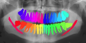
- 2D Teeth Modeling
Each tooth is assigned a unique color, allowing for the tracking of individual teeth over time. In dental surgery, this segmentation is crucial as it encourages the surgeon to plan the procedure based on the position and alignment of the teeth. Read more in my blog.
- 3D Teeth Modeling
This image displays 3D Intraoral Surface (IOS) scans of teeth with automatic labeling.